
In our lab we use Drosophila melanogaster to model several types of cancer with the hopes of finding new molecular pathways that can be targeted in cancer therapy. This generates a huge amount of data, some of which needs impartial analysis, which is where you could help!
Project description
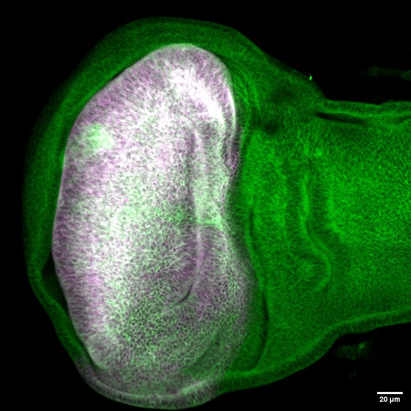
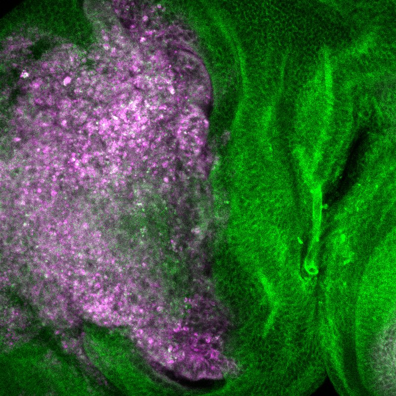
I am using Drosophila to model cancers with Liver Kinase B1 (LKB1) mutation. LKB1 is a contextual oncogene, meaning both inactivation (tumour suppressive role) and over activation (oncogenic role) can make a cancer more aggressive, resulting in poor patient prognosis. I have induced cancer in several fly lines and see subtle but clear differences depending on the level of LKB1 present. To avoid any bias I would like a student to complete a qualitative analysis of the images with help from ImageJ. This will then generate data which can be analysed in R (depth of analysis dependent on the amount of time the student has remaining).
Tasks
The student will be given their own set of images to work with. This will be quite large, so they will need to remain organised so the data output remains clear. They will analyse the images using ImageJ, and input data into Excel. I hope there is then time for the student to also learn the basics of R to analyse the data they have generated, and create plots alongside this to visualise it.
Experience
No previous experience is required 🙂 However, a laptop/computer that can run ImageJ and R is necessary.
Students will learn to use ImageJ and hopefully the basics of R!
They will also receive valuable experience on how to organise large data sets in Excel.
Starting date/period: 01 Feb 2026 – 30 Apr 2026
The project involves: computer work
Involvement: 40 hours, but the student can pick their own working hours as not lab based.
Interested by this project? Need more info? Contact Katherine Downham (klp.downham@gmail.com)
Project number: 055